New publication in Analytical Chemistry!
First publication date: 01/06/2020

Congratulations Maite and Alba for you new publication in Analytical Chemistry!
Thanks to all contributors! It is a great piece of work carried out by Maite Garcia-Hernando, Alba Calatayud-Sanchez; Jaione Etxebarria-Elezgarai; Marian M de Pancorbo, Fernando Benito-Lopez and Lourdes Basabe-Desmonts, entitled: Single Cell Resolution Cytotoxicity Biosensor Based on Single Cell Adhesion Dot Arrays.
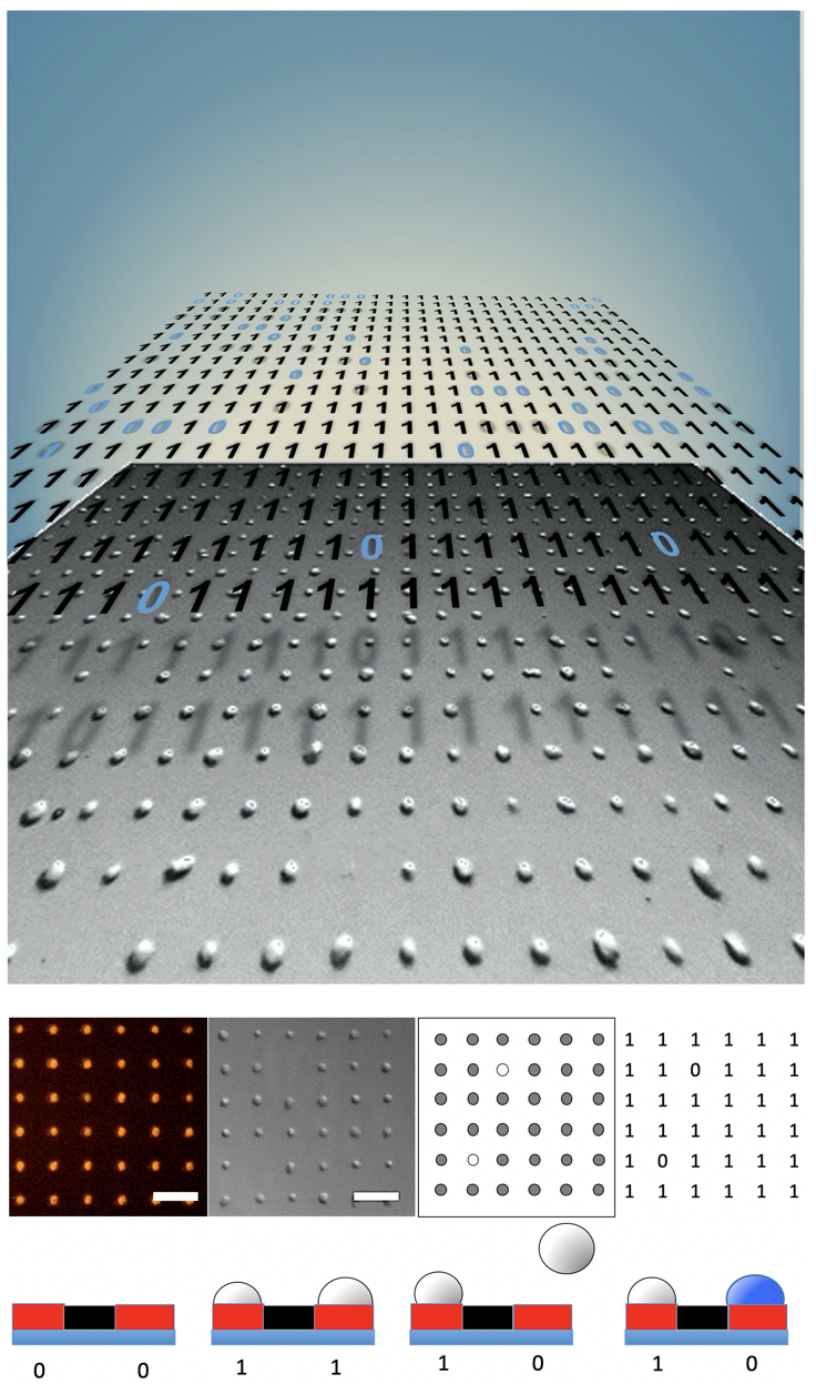
The manuscript presents an optical biosensor platform for dynamic measurements of cell cytotoxicity with single cell resolution. Cell viability tests are needed in the pharmaceutical, biomaterial and enviromental industry to measure adverse cellular effects. Available cytotoxicity tests, are highly time consuming and require trained personal and expensive labels and instrumentation. "Single Cell Adhesion Dot Arrays" (SCADA), enabled digital cytometric quantification of cell viability by simple optical imaging. Fibronectin dots arrays were fabricated by microcontact printing on cell culture multiwells plates. The dot array was designed to allocate a single cell in each fibronectin dot. Digital dynamic monitoring of cell adhesion was performed by quantifying the number of occupied dots by optical imaging without the need of labels. For cytotoxicity measurements cells-filled SCADA substrates were exposed to K2CrO4, HgSO4 salts and dimethyl sulfoxide (DMSO). Dynamic monitoring of the toxic effect of DMSO and K2CrO4 was done measuring cell detachment rate during more than 30 hours. HgSO4 inhibited cellular detachment from the surface, and cytotoxicity was monitored using Trypan Blue life/death assay directly on the surface. In all cases the cytotoxicity effects were easily monitored and comparable to previous reports. These assays require only a transparent substrate area of less than 1 mm2 and a reduced number of cells, it could be integrated into microfluidic platforms to provide a practical tool simple to use. This methodology for accurate measurement of cell adhesion, detachment and staining could extent to fundamental research and commercial applications.
Links

