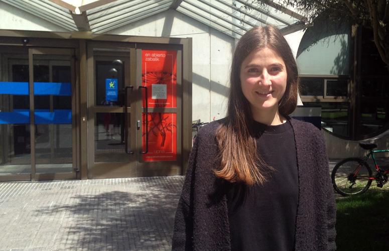
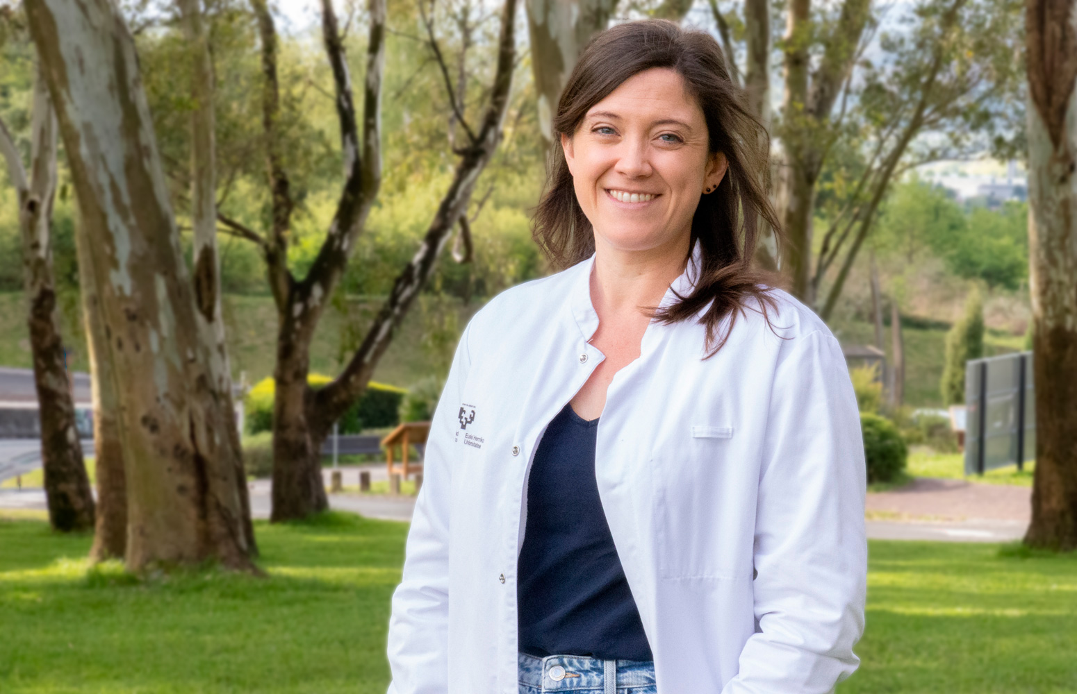
At the Auckland Institute of Technology (New Zealand) the UPV/EHU’s Biomat group used a 3D printer to produce gelatin scaffolds to assist in tissue regeneration. The printer allows the scaffolds to be adapted to the exact needs of the patient and their composition may also include the necessary components. In this research, for example, dexamethasone was added as an anti-inflammatory drug and lactose as a crosslinking agent to provide the biomaterial with greater stability in aqueous environments.
Gelatin scaffolds personalised in terms of shape and composition using 3D printers
The Biomat group of the UPV/EHU-University of the Basque Country has created biomaterials at the Auckland Institute of Technology using gelatin that is a by-product of industrial waste
- Research
First publication date: 16/05/2019

In regenerative medicine there is a growing trend for developing biodegradable materials to help tissue to regenerate. Advanced manufacturing techniques such as 3D printers are also being used to assist in this direction. The BIOMAT-biopolymeric materials research group attached to the UPV/EHU’s Faculty of Engineering - Gipuzkoa combined both of them and designed a gelatin biomaterial. In other words, they used a 3D printer to produce a scaffold that could be used in regenerative medicine. “What is more, our raw material was gelatin that is a by-product of meat industry waste, so we were also able to upgrade waste,” explained Alaitz Etxabide, a member of the BIOMAT research group and lead author of the paper.
3D printers offer the chance to adapt both the design and the composition of the scaffold to the needs of each patient, “which current standard scaffolds do not do. Scaffolds can be designed on the basis of the individual anatomy of each patient by using specific software for this purpose so that adaptation is absolute”, explained the doctor in renewable materials engineering.
The “toner” designed for use in the 3D printer was a blend of a hydrogel type, the main component of which was the gelatin obtained by means of the hydrolysis of the above-mentioned collagen waste. “The main drawback we had to address was that gelatin dissolves very fast in aqueous environments, and even faster at temperatures such as that of the body,” pointed out Etxabide. And even though it is true that it is preferable for scaffolds for use in biomedicine to be dissolved and digested by the organism, it is advisable for this to take place after some time, the time needed for the damaged tissue to regenerate. “So we added lactose to the blend believing that it would slow down the degradation of the gelatin when reacting with it,” she said.
Apart from the gelatin and lactose, a third component was included in the designed toner: dexamethasone, an anti-inflammatory, immunosuppressive drug. “If the scaffold itself that is placed in the damaged tissue releases a drug that assists healing, a smaller dose of the drug will be needed to obtain the same effect and, what is more, the effect will be achieved faster, because it is not necessary to wait until it has been digested by the organism, as happens with drugs administered by mouth,” she added. As the researcher highlighted, “it is possible to tweak the formulation of the toner, and add some components or others to achieve the desired effect in the patient”.
Once the scaffold had been designed and its formulation specified, the third step was to print it. “In our research we opted for liquid toner because it offers greater versatility for tweaking the formulation than materials in solid or powder form. However, we had to bear in mind that the liquid needed to have a minimum viscosity so that it would keep its shape once printed”, said Etxabide. After leaving it at room temperature for 24 hours, the water gradually evaporated and finally a totally solid structure was achieved. Afterwards, it was subjected to a heating process to encourage the gelatin and the lactose to react with each other and render the scaffold more resistant in the presence of water.
The fourth and final step of the process would be to insert the manufactured scaffold into the damaged organism. The researcher is aware of the long journey that lies ahead "until the scaffolds can begin to be used with actual patients, but in the study we simulated the conditions of the human body, as we put the scaffold in conditions of temperature and humidity typical to those of the body to study how it would evolve inside the organism, since our research was aiming to test whether it would be possible to produce a scaffold suitable for use in biomedicine. It was a pre-analysis”. And the results of the analysis showed that the reaction with the lactose extended the duration of the material by up to seven days, otherwise the material would have dissolved in water within a day. Furthermore “we saw that the surface of the material was rough, which makes in particularly suited for use as a scaffold, given that this helps the cells to become adhered to it", specified the researcher.
Following the pre-analysis they are continuing to work along the long road towards the final aim of using the scaffold in medicine. Etxabide explained, “We are collaborating with another group at the UPV/EHU as our group does not have the capacity to conduct the in vitro and in vivo studies that would be needed; in our group we work on the properties of materials and on production methods, but we need to work with research groups from other areas of expertise so that each one can make its own contributions”.
Additional information
A research grant enabled Alaitz Etxabide, researcher in the BIOMAT-biopolymeric materials research group of the UPV/EHU’s Faculty of Engineering - Gipuzkoa, to conduct this study at the Auckland Institute of Technology, New Zealand. In her study Etxabide made use of the 3D printer belonging to that institute.
Bibliographical reference
Alaitz Etxabide, Jingjunjiao Long, Pedro Guerrero, Koro de la Caba, Ali Seyfoddin
3D printed lactose-crosslinked gelatin scaffolds as a drug delivery system for dexamethasone.
European Polymer Journal (2019)


