

Temario y Bibliografía
- La ilustración médica es la rama del dibujo científico que se dedica a la anatomía y organografía humana, a la anatomía quirúrgica y a ilustrar los procesos fisiológicos y patológicos del organismo humano; también participa en la presentación de datos clínicos e infografías relacionadas con la actividad médica.
- 1. Anatomía macroscópica
- 1.1. Terminología básica: posición anatómica, planos de sección e indicaciones topográficas. Regiones y sistemas.
- 1.2. Errores frecuentes de la ilustración anatómica.
- 1.3. Dibujo musculoesquelético: formas y texturas de huesos, articulaciones, tendones y músculos.
- 1.4. Dibujo visceral: formas y texturas de vasos sanguíneos y órganos abdómino-torácicos.
- 1.5. Dibujo neuroanatómico: particularidades del sistema nervioso central y órganos de los sentidos.
- 2. Anatomía microscópica
- 2.1. Terminología histológica.
- 2.2. La célula: estructura e ilustración de sus componentes.
- 2.3. Los cuatro grandes grupos tisulares: tejido epitelial, tejido conectivo, tejido muscular y tejido nervioso.
- 2.4. Ilustración de anatomía microscópica de diversos órganos.
- 3. Ilustración quirúrgica y médica aplicada
- 3.1. El quirófano: elementos físicos y normas, medidas de asepsia, ¿cómo comportarse dentro de un quirófano?
- 3.2. Técnicas quirúrgicas básicas: campo quirúrgico, incisiones, disecciones, suturas, instrumental básico.
- 3.3. Ilustrar pasos de una cirugía.
- 3.4. Ilustrar la enfermedad: los grandes síndromes, signos y síntomas, lesiones y maniobras de exploración.
- 4. Infografía médica
- 4.1. Ilustración de procesos fisiológicos y fisiopatológicos.
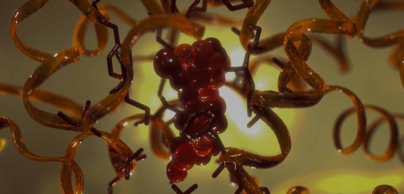
- 4.2. Ilustración molecular y farmacológica.
- 4.3. La infografía aplicada a la presentación de datos clínicos.
Bibliografía:Illustrated Encyclopedia of Human Histology. RV Krstic. Springer, 1984.
Bibliografía:Illustrated Encyclopedia of Human Histology. RV Krstic. Springer, 1984.
Bibliografía:Illustrated Encyclopedia of Human Histology. RV Krstic. Springer, 1984.
Bibliografía:Illustrated Encyclopedia of Human Histology. RV Krstic. Springer, 1984.
Bibliografía:Illustrated Encyclopedia of Human Histology. RV Krstic. Springer, 1984.
Bibliografía:Illustrated Encyclopedia of Human Histology. RV Krstic. Springer, 1984.
Bibliografía:Illustrated Encyclopedia of Human Histology. RV Krstic. Springer, 1984.
Bibliografía:Illustrated Encyclopedia of Human Histology. RV Krstic. Springer, 1984.
Bibliografía:Illustrated Encyclopedia of Human Histology. RV Krstic. Springer, 1984.
Bibliografía:Illustrated Encyclopedia of Human Histology. RV Krstic. Springer, 1984.
Bibliografía:Illustrated Encyclopedia of Human Histology. RV Krstic. Springer, 1984.
Bibliografía:Illustrated Encyclopedia of Human Histology. RV Krstic. Springer, 1984.
Bibliografía:Illustrated Encyclopedia of Human Histology. RV Krstic. Springer, 1984.
Bibliografía:Illustrated Encyclopedia of Human Histology. RV Krstic. Springer, 1984.
Bibliografía:Illustrated Encyclopedia of Human Histology. RV Krstic. Springer, 1984.
Bibliografía:Illustrated Encyclopedia of Human Histology. RV Krstic. Springer, 1984.
Bibliografía:Illustrated Encyclopedia of Human Histology. RV Krstic. Springer, 1984.
Bibliografía:Illustrated Encyclopedia of Human Histology. RV Krstic. Springer, 1984.
Bibliografía:Illustrated Encyclopedia of Human Histology. RV Krstic. Springer, 1984.
Bibliografía:Illustrated Encyclopedia of Human Histology. RV Krstic. Springer, 1984.
Bibliografía:¿Gray: Anatomía para estudiantes. R.L. Drake. Elsevier, 2014.